Day 1 Monday, February 25, 2019
8:00 Light Breakfast and Registration
8:50 Welcoming Remarks/Convener David Andrews
Education Session 1:
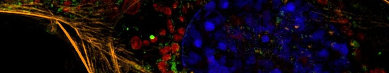
9:00 Imaging Mass Cytometry: High-dimension Imaging, Segmentation and Analysis of Spatially Resolved Single Cells
Hartland Jackson, Institute for Molecular Life Sciences, University of Zürich
9:45 Multiplexed Imaging for Multi-protein Analysis at the Single Cell Level in Fixed Tissue (MultiOmyx™ and Cell DIVE™) Fiona Ginty, Biosciences Technology Manager & Principal Investigator at GE Global Research
10:30 3D Models for High Content Analysis
Judi Wardwell, InSphero
11:15 3D Models for Cancer Biology
Shannon Mumenthaler, University of Southern California
12:00-1:30 Lunch – Installation of Cell Profiler and dataset – bring your laptop.
Education Session 2:
Convener: Mike Mancini
1:30 Rapid Quantification of Protein-protein Interactions in Live Cells
David Andrews, University of Toronto
2:15 Statistical Analysis of High Content Screening Data
Bartek Rajwa, Purdue University
3:15 Break
3:30-5:00 Cell Profiler Workshop
Santosh Hariharan, Pfizer
Bring your own laptop with Cell Profiler installed.
Day 2 Tuesday, February 26, 2019
8:30 Light Breakfast and Registration
9:00 Welcoming Remarks
Mike Mancini, Baylor College of Medicine
David Andrews, University of Toronto
Session 1: Nuclear Genomics and Chromatin Organization
Convener: Mike Mancini, Baylor College of Medicine
9:15 Keynote Presentation:
Spatial Genomics: Nascent Transcriptome Profiling by intron seqFISH.
Long Cai, California Institute for Technology
10:00 3D HCS Imaging Challenges and Solutions for Complex Cell Model Systems
Erez Leiberman, Baylor College of Medicine
10:30 Break
10:45 Microscopic Imaging of Epigenetic Landscape in Single Cells
Alexey Terkskikh, Ph.D., Sanford Burnham Prebys Research Institute
11:15 Nuclear Speckles as a Transcriptional Hub and Amplifier: A Combined Genomic and Live-cell Imaging Approach
Andrew Belmont, University of Illinois at Urbana-Champaign
11:45 Selected Abstract:
Imaging Freeze-frame Proteins Identifies Cancer-Protein Functions and Protein Networks of Endogenous DNA Damage
Jun Xia, Baylor College of Medicine
12:00 Lunch and Poster Session
12:45 Presenters at Posters
Session 2: Advanced Cellular Models
Convener: Fabio Stossi, Baylor College of Medicine
1:30 Non-opsin Based Optogenetics to Illuminate Physiology
Yubin Zhou, Institute of Biosciences and Technology, Texas A&M Health Science Center
2:00 The Single Cell Pathology Landscape of Breast Cancer: Tumour Cells and Their Microenvironments
Hartland Jackson, Institute for Molecular Life Sciences, University of Zürich
2:30 Multiplex Imaging for Single Cell Mechanistic Analysis of Estrogen Receptor Functions
Mancini/Stossi, Baylor College of Medicine
3:00 Break
3:15 Expansion Microscopy: A novel tool for Single Cell Analysis in Intact Biological Systems
Mahander Dewal, Expansion Technologies, Inc.
3:45 Selected Abstract:
A 3D in vitro Platform for High-Throughput Screening of Diverse Prostate Cancer Patient-Derived Xenograft Models
Lindsey Sablatura, Rice University
Day 3 Wednesday, February 27, 2019
8:00 Light Breakfast and Registration
Session 3: Image Informatics
Convener: David Andrews, University of Toronto
9:00 Keynote Presentation:
Integrating Information from Diverse Microscope Images: Learning and Using Generative Models of Cell Organization
Bob Murphy, Carnegie-Mellon University
9:45 Phindr3D: Data-driven Segmentation-free Phenotyping of 3D Santosh Hariharan, Pfizer
10:15 Spheroid Imaging and Sequencing
Christian Conrad, Berlin Institute of Health, Germany
10:45 Break
11:00 OME’s Bio-Formats, OMERO, & IDR: Open Tools for Accessing, Integrating, Mining and Publishing Image Data @ Scale
Jason Swedlow, University of Dundee
11:30 Magnetic 3D Bioprinting, from Generating Spheroids to Fingerprinting Cell-Types
Glauco Souza, Greiner Bio One
12:00 Bridging the Phenotypic-Genomic Continuum: Case Studies Linking Histology and Genomics
Arvind Rao, University of Michigan
12:30 Lunch and Posters
Session 4: Image-Based Assays for Cancer Research
Convener: Peter Davies, Institute of Biosciences and Technology, Texas A&M Health Science Center
2:00 Image-based Assays Reveal Fibroblast-Mediated Drug Resistance in Colorectal Cancer
Shannon Mumenthaler, University of Southern California
2:30 High Content Chemoresponse Assays for Personalized Management of Cancer
David Andrews, University of Toronto
3:00 Selected Abstract:
3D Modeling Of Chromosomes Territories in Normal and Aneuploid Nuclei
Fatima Merchant, University of Houston
3:15 3D HCS Imaging Challenges and Solutions for Complex Cell Model Systems
Joe Trask, Perkin Elmer
3:45 Break
4:00 Fluorescent multiplex IHC: An integrated approach for high throughput panel- driven and ultra-high-plex multiplexing tissue with single-cell resolution
Grady Carlson, Akoya Biosciences
4:30 Protein Marker Multiplexing and Quantitative Image Analysis for Disease Characterization
Alison Cheung, Sunnybrook Research Institute